Elements of Visual Perception
Also known as the Structure of the human Eye. The eye is nearly a sphere with average approximately 20 mm diameter. The eye is enclosed with three membranes.
1. Cornea and Sclera
it is a tough, transparent tissue that covers the anterior surface of the eye. Rest of the optic globe is covered by the sclera.
2. Choroid
It contains a network of blood vessels that serve as the major source of nutrition to the eyes. It helps to reduce extraneous light entering in the eye It has two parts
- Iris Diaphragms: it contracts or expands to control the amount of light that enters the eyes.
- Ciliary body
3. Retina
it is innermost membrane of the eye. When the eye is properly focused, light from an object outside the eye is imaged on the retina. There are various light receptors over the surface of the retina.
- Cones: it is in the number about 6 to 7 million. These are located in the central portion of the retina called the fovea. These are highly sensitive to color. Human can resolve fine details with these cones because each one is connected to its own nerve end. Cone vision is called photopic or bright light vision.
- Rods: These are very much in number from 75 to 150 million and are distributed over the entire retinal surface. The large area of distribution and the fact that several roads are connected to a single nerve give a general overall picture of the field of view. They are not involved in the color vision and are sensitive to low level of illumination. Rod vision is called is scotopic or dim light vision.

The absent of reciprocators is called blind spot.
Image Formation in the Eye
The major difference between the lens of the eye and an ordinary optical lens in that the former is flexible.
The shape of the lens of the eye is controlled by tension in the fiber of the ciliary body. To focus on the distant object the controlling muscles allow the lens to become thicker in order to focus on object near the eye it becomes relatively flattened.
The distance between the center of the lens and the retina is called the focal length and it varies from 17mm to 14mm as the refractive power of the lens increases from its minimum to its maximum.
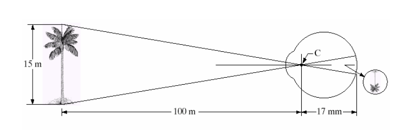
When the eye focuses on an object farther away than about 3m. The lens exhibits its lowest refractive power. When the eye focuses on a nearly object. The lens is most strongly refractive.
The retinal image is reflected primarily in the area of the fovea. Perception then takes place by the relative excitation of light receptors, which transform radiant energy into electrical impulses that are ultimately decoded by the brain.
Brightness Adaption and Discrimination
Digital image are displayed as a discrete set of intensities. The range of light intensity levels to which the human visual system can adopt is enormous- on the order of 1010 from scotopic threshold to the glare limit.
Experimental evidences indicate that subjective brightness is a logarithmic function of the light intensity incident on the eye.
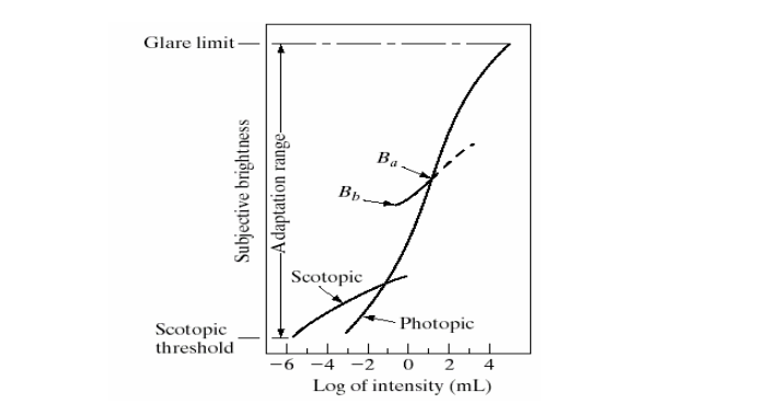
The curve represents the range of intensities to which the visual system can adopt. But the visual system cannot operate over such a dynamic range simultaneously. Rather, it is accomplished by change in its overcall sensitivity called brightness adaptation.
For any given set of conditions, the current sensitivity level to which of the visual system is called brightness adoption level , Ba in the curve. The small intersecting curve represents the range of subjective brightness that the eye can perceive when adapted to this level.
It is restricted at level Bb , at and below which all stimuli are perceived as indistinguishable blacks. The upper portion of the curve is not actually restricted. whole simply raise the adaptation level higher than Ba.
The ability of the eye to discriminate between change in light intensity at any specific adaptation level is also of considerable interest.